In Vivo and Ex Vivo Services to Expedite Preclinical Testing
7 April 2023
About Pharmacology Discovery Services (PDS)
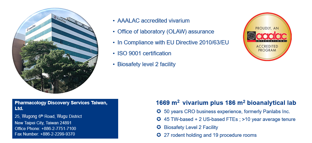
Pharmacology Discovery Services (PDS), a partner lab of Eurofins Discovery, provides a comprehensive collection of validated in vivo oncology models to present key decision-making information for allowing test agents to move forward. Our services provide the data clients need to advance their preclinical development programs in a timely, flexible, and cost-effective manner. Each model can be customized to meet specific pharmacology needs.
Our in vivo oncology models include mouse tumor allografts (syngeneic models) and human xenografts from both cell line-derived (CDX) and patient-derived (PDX) samples. To assess new testing agents for their ability to promote anti-cancer responses, our CDX and PDX models, including subcutaneous and orthotopic formats, have been developed and validated for responses to known standard-of-care treatments. Thus, these models are suitable for the evaluation of efficacious treatment followed by ex vivo analysis with different platforms including flow cytometry, RT-qPCR, ELISA, western blot, histopathology, and bioanalysis.
The carcinoma cell lines used for our CDX models are accessible from global cell banks, including the American Type Culture Collection (ATCC), the Japanese Collection of Research Bioresources Cell Bank (JCRB), and the RIKEN BRC Cell Bank. Through our Partner Lab, Eurofins Discovery, in vitro screening with these cell lines is available with OncoPanel™ cell-based screening and profiling, as well as test agents and combinations with BioMAP® Oncology and Immune Oncology Panels in modeling the human tumor microenvironment.
PDS has conducted in vivo pharmacology studies for both side-effect profiling and efficacy for over 50 years. We have the flexibility and expertise to develop custom models and are experienced with small molecules, drug combinations, biologics, nucleic acids, liposomal formulations, nanoparticles, and vaccines.
Overview of in Vivo Oncology Services
In Vivo Oncology Model Types
- Murine cell line-derived syngeneic models with immune-competent mice
- Human cell line-derived xenograft models with immune-deficient mice
- Human patient-derived xenograft models with immune-deficient mice
- Humanized mouse models with greater than 30% hCD45+ cells
- - Models are available with subcutaneous and orthotopic implantation
- - Site-specific tumor implantation for metastatic evaluation is also available
Optimal Measurement Readouts Upon Selection
- Conventional caliper tumor measurements
- Conventional macroscopic metastatic nodules
- In vivo imaging with IVIS® Spectrum
- Survival analysis with humane endpoints
- Flow cytometry analysis with leukemia/lymphoma progression
- Histopathology, cytokine, flow cytometry, RT-qPCR, ELISA, and western blot
- Pharmacokinetics (PK) bioanalysis
Flexible Dosing Routes and Schedules
- Administration routes include but not limited to: oral (PO), subcutaneous (SC), intraperitoneal (IP), intravenous (IV), and intratumoral (Itu) injections
- Dosing frequencies include single or multiple doses per day (QD, BID, TID, and QID), per week (QWK, BIW, and TIW), or at a specific number of days for on and off dosing (i.e. 3 on/4 off per week)
- Formulation optimization services with customized vehicles
- Dose concentration and frequency optimizations with MTD and PK studies
Customized Models and Extensive Ex Vivo Assays to Tailor Pharmacology Needs
- - Collaborate with our technical staff to develop models specific to your needs
- - Extensive ex vivo assays available as optional services for flexible selection upon review of weekly interim data.
- - Experienced in cell labeling and genetically engineering of cell lines.
Our portfolio is expanding rapidly. To get started, speak to our expert: PanlabsTaipeiLab@eurofinsus.com for availability in your model of interest.

In Vivo Syngeneic Mouse Models
Syngeneic mouse models with immune-competent mice provide a comprehensive effective approach for in vivo efficacy studies. Our syngeneic mouse models (Table 1) include subcutaneous and orthotopic formats, and have been developed and validated for responses to known chemotherapy and immunotherapy treatments, including immune checkpoint inhibitors. Analysis of tumor infiltrating leukocytes is conducted by flow cytometry and is available with different models.
| Validated Syngeneic Models | |||
|---|---|---|---|
| Model Name | Item Number | Cell Lines Used in Mouse Model | |
| Tumor, Syngeneic, Breast, 4T1 | 578590 | ATCC | CRL-2539 |
| Tumor, Syngeneic, Colon, CT26.WT | 578600 | ATCC | CRL-2638 |
| Tumor, Syngeneic, Colon, MC-38 | 578620 | Keradast | ENH204-FP |
| Tumor, Syngeneic, Kidney, Renca | 578630 | ATCC | CRL-2947 |
| Tumor, Syngeneic, Leukemia, L1210 | 578650 | ATCC | CRL-219 |
| Tumor, Syngeneic, Lung, KLN 205 | 578680 | ATCC | CRL-1453 |
| Tumor, Syngeneic, Lung, LL/2 | 578700 | ATCC | CRL-1642 |
| Tumor, Syngeneic, Melanoma, B16-F0 | 578800 | ATCC | CRL-6322 |
| Tumor, Syngeneic, Melanoma, B16-F10 | 578810 | ATCC | CRL-6475 |
| Tumor, Syngeneic, Lymphoma, A20 | 578900 | ATCC | TIB-208 |
Table 1. Validated syngeneic models.
In Vivo Xenograft Mouse Models
Xenograft mouse models with human cells play an important role in the screening and evaluation of novel anti-cancer test agents. The models can be derived from human tumor cell lines (CDX models) or patient tumors (PDX models) and are utilized to evaluate therapeutic efficacy and toxicity. The tumor cells are implanted subcutaneously (Table 2) or orthotopically (Table 3) in immune-deficient mice or mice with human immune cells (humanized mice). The therapeutic profile with standard-of-care drugs can be provided upon request for each model. Analysis of tumor infiltration is conducted by flow cytometry and is available with different tissues.
| Xenograft Models | |||
|---|---|---|---|
| Model Name | Item Number | Cell Lines used in mouse model | |
| Tumor, Xenograft, Brain, U-87 MG | 579500 | ATCC | HTB-14 |
| Tumor, Xenograft, Breast, BT-474 | 579700 | ATCC | HTB-20 |
| Tumor, Xenograft, Breast, HCC1428 | 579800 | ATCC | CRL-2327 |
| Tumor, Xenograft, Breast, JIMT-1 | 579900 | AddexBio | C0006005 |
| Tumor, Xenograft, Breast, MCF7 | 580000 | ATCC | HTB-22 |
| Tumor, Xenograft, Breast, MDA-MB-231 | 580020 | ATCC | HTB-26 |
| Tumor, Xenograft, Breast, T-47D | 580030 | ATCC | HTB-133 |
| Tumor, Xenograft, Bone, U-2 OS | 580080 | ATCC | HTB-96 |
| Tumor, Xenograft, Bladder, UM-UC-3 | 580500 | ATCC | CRL-1749 |
| Tumor, Xenograft, Colon, HCT 116 | 580090 | ATCC | CCL-247 |
| Tumor, Xenograft, Colon, HT-29 | 580100 | ATCC | HTB-38 |
| Tumor, Xenograft, Colon, SW480 | 580150 | ATCC | CCL-228 |
| Tumor, Xenograft, Head/Neck, SAS | 580300 | JCRB | JCRB0260 |
| Tumor, Xenograft, Kidney, A-498 | 580400 | ATCC | HTB-44 |
| Tumor, Xenograft, Kidney, ACHN | 580450 | ATCC | CRL-1611 |
| Tumor, Xenograft, Leukemia (PML/AML/M2), HL-60 | 580600 | ATCC | CCL-240 |
| Tumor, Xenograft, Leukemia (AML/M5), MV-4-11 | 580650 | ATCC | CRL-9591 |
| Tumor, Xenograft, MDS/Leukemia (AML/M5), SKM-1 | 580670 | JCRB | JCRB0118 |
| Tumor, Xenograft, Leukemia (AMoL/M5), THP-1 | 580680 | ATCC | TIB-202 |
| Tumor, Xenograft, Liver, Hep 3B2.1-7 | 580700 | ATCC | HB-8064 |
| Tumor, Xenograft, Liver, PLC/PRF/5 | 580730 | ATCC | CRL-8024 |
| Tumor, Xenograft, Lung (NSCLC), A549 | 580900 | ATCC | CCL-185 |
| Tumor, Xenograft, Lung (NSCLC), NCI-H1975 | 580910 | ATCC | CRL-5908 |
| Tumor, Xenograft, Lung (SCLC), NCI-H209 | 580940 | ATCC | HTB-172 |
| Tumor, Xenograft, Lung (NSCLC), NCI-H460 | 580950 | ATCC | HTB-177 |
| Tumor, Xenograft, Lung (SCLC), NCI-H526 | 580960 | ATCC | CRL-5811 |
| Tumor, Xenograft, Lung (NSCLC), PC-9 | 580970 | Sigma Aldrich | 90071810 |
| Tumor, Xenograft, Lymphoma (AML/M5), U-937 | 581000 | ATCC | CRL-1593.2 |
| Tumor, Xenograft, Lymphoma, SU-DHL-5 | 581010 | ATCC | CRL-2958 |
| Tumor, Xenograft, Neuroepithelioma (Askin), SK-N-MC | 581100 | ATCC | HTB-10 |
| Tumor, Xenograft, Ovary, SK-OV-3 | 581300 | ATCC | HTB-77 |
| Tumor, Xenograft, Ovary, OVCAR-3 | 581330 | ATCC | HTB-161 |
| Tumor, Xenograft, Pancreas, MIA PaCa-2 | 581500 | ATCC | CRL-1420 |
| Tumor, Xenograft, Pancreas, BxPC-3 | 581520 | ATCC | CRL-1687 |
| Tumor, Xenograft, Pancreas, PANC-1 | 581540 | ATCC | CRL-1469 |
| Tumor, Xenograft, Prostate, PC-3 | 581900 | ATCC | CRL-1435 |
| Tumor, Xenograft, Prostate, 22Rv1 | 581920 | ATCC | CRL-2505 |
| Tumor, Xenograft, Prostate, DU 145 | 581930 | ATCC | HTB-81 |
| Tumor, Xenograft, Prostate, LNCaP clone FGC | 581950 | ATCC | CRL-1740 |
| Tumor, Xenograft, Skin, A-431 | 582000 | ATCC | CRL-1555 |
| Tumor, Xenograft, Melanoma, A-375 | 582100 | ATCC | CRL-1619 |
| Tumor, Xenograft, Melanoma, SK-MEL-5 | 582110 | ATCC | HTB-70 |
| Tumor, Xenograft, Myeloma, RPMI 8226 | 582200 | ATCC | CCL-155 |
| Tumor, Xenograft, Neuroblastoma, IMR-32 | 582300 | ATCC | CCL-127 |
| Tumor, Xenograft, Neuroblastoma, SK-N-AS | 582310 | ATCC | CRL-2137 |
| Tumor, Xenograft, Gastric, NCI-N87 | 582400 | ATCC | CRL-5822 |
Table 2. Validated cell line derived xenografts (CDX).
In Vivo Orthotopic Mouse Models
Orthotopic mouse models with murine and human cells play an important role in assessing tumor development in a relevant environment to evaluate the efficacy of therapeutic treatment that mimics the disease process in humans. The tumor cells are orthotopically implanted in the tissue of carcinoma origin. These models (Table 3) include the implantation of breast tumor cells into the mammary fat pad, pancreatic tumor cells into the pancreas, and by tail vein injection with leukemic cells (also known as disseminated models). Tumor cells that have metastatic potential will result in spontaneous metastasis that mimics the human disease progression as demonstrated with the highly metastatic TNBC murine 4T1 and human MDA-MB-231 orthotopic models with. In addition to the tracking of tumor cells with IVIS® Spectrum, flow cytometry can be applied for monitoring leukemic cells in the bloodstream. If metastasis to a specific organ is of interest, our team is experienced with implantation via different injection routes, including directly into the pancreas, portal vein injection to reach liver tissues, and intracardiac injection for brain metastasis.
Patient-Derived Xenografts (PDX Models)
Patient-derived tumor xenografts are developed by directly transplanting tumor fragments from surgically dissected cancer patients into immunodeficient or humanized mice. The model is useful for translational research by retention of the original tumor architecture and facilitating precision medicine. The susceptibility of a therapeutic compound that results in tumor reduction in PDX models is closely correlated with clinical data in patients from whom the PDX models were derived. This suggests that PDX models are highly effective in predicting the efficacy of both conventional and novel anti-cancer therapeutics.
| Orthotopic Models | |||
|---|---|---|---|
| Model Name | Item Number | Cell Lines used in mouse model | |
| Tumor, Orthotopic, Breast, 4T1 | 578591 | ATCC | CRL-2539 |
| Tumor, Orthotopic, Breast, 4T1-Luc* | 578592 | JCRB | JCRB1447 |
| Tumor, Orthotopic, Breast, KLN 205-Luc/GFP* | 578682 | ATCC | CRL-1453, and then labeled by PDS# |
| Tumor, Orthotopic, Brain, U-87 MG-Luc* | 579502 | PerkinElmer | BW124577 |
| Tumor, Orthotopic, Breast, MDA-MB-231 | 580021 | ATCC | HTB-26 |
| Tumor, Orthotopic, Breast, MDA-MB-231-Luc* | 580022 | JCRB | JCRB1559 |
| Tumor, Orthotopic, Leukemia (PML/AML/M2), HL-60 | 580601 | ATCC | CCL-240 |
| Tumor, Orthotopic, Leukemia (AML/M5), MV-4-11 | 580651 | ATCC | CRL-9591 |
| Tumor, Orthotopic, Lymphoma, U-937 | 581001 | ATCC | CRL-1593.2 |
| Tumor, Orthotopic, Pancreas, MIA PaCa-2 | 581501 | ATCC | CRL-1420 |
| Tumor, Orthotopic, Pancreas, BxPC-3 | 581521 | ATCC | CRL-1687 |
| Tumor, Orthotopic, Pancreas, BxPC-3-Luc* | 581522 | PerkinElmer | BW125058 |
| Tumor, Orthotopic, Pancreas, PANC-1 | 581541 | ATCC | CRL-1469 |
Table 3. Cell line derived orthotopic models.
* Denotes the cell line is labeled with luciferase for monitoring with IVIS Spectrum.
# Denotes the cell line was labeled by PDS using lenti-viral particles followed by selection.
| PDX Models | |||
|---|---|---|---|
| Model Name | Item Number | Specimen information | |
| Tumor, Colon, PDX, T18 | 585100 | KRAS mutation | Stage IV A |
| Tumor, Pancreas, PDX, (coming soon, available in ASID) | |||
| Tumor, Gastric, PDX, (coming soon, available in ASID) | |||
Table 4. Patient-derived models.
New model development and ex vivo analysis
Pharmacology Discovery Services is very experienced with co-development of new models. These models are owned by our Sponsors and although cannot be offered as a service, the knowledge for successful model application can be provided. Pharmacology Discovery Services provides a full-service facility equipped for in vitro, in vivo, and ex vivo analyses. Supplemental ex vivo analysis during in vivo studies is often an important components of a complete preclinical program. These ex vivo analyses are important for understanding the function and mechanism of the in vivo effect of a test article, as well as for exploring the correlation between in vivo efficacy and biomarkers.