
Lupus, Systemic Lupus Erythematosus (SLE), MRL/lpr Mouse
CONTACT CUSTOMER SERVICE
ADD TO FAVORITES
ADD TO PANEL
OVERVIEW
DOCUMENTS/REFERENCES
BIOLOGICAL INFORMATION
BACKGROUND INFORMATION
Systemic lupus erythematosus (SLE) is an autoimmune disease that is characterized by autoantibody production and involves many organs such as skin, joints, kidneys, and the central nervous system (CNS), and the renal disease (lupus nephritis) is considered the most severe manifestation of SLE with an increased risk of morbidity and mortality. The two most commonly used spontaneous mouse models are MRL/lpr mice and NZBWF1/J mice. MRL/lpr mice are homozygous for spontaneous mutation in the Fas gene and develop enlarged lymph nodes and spleen, anti-nuclear and anti-dsDNA antibodies, and systemic autoimmunity. The MRL/lpr mouse model has been used extensively to study the mechanisms of SLE and evaluate the therapeutic efficacy of drug candidates.
ORGANISM
Mouse
ASSAY INFORMATION
ASSAY TYPE
In Vivo
TESTING INFORMATION
PROCEDURE SUMMARY
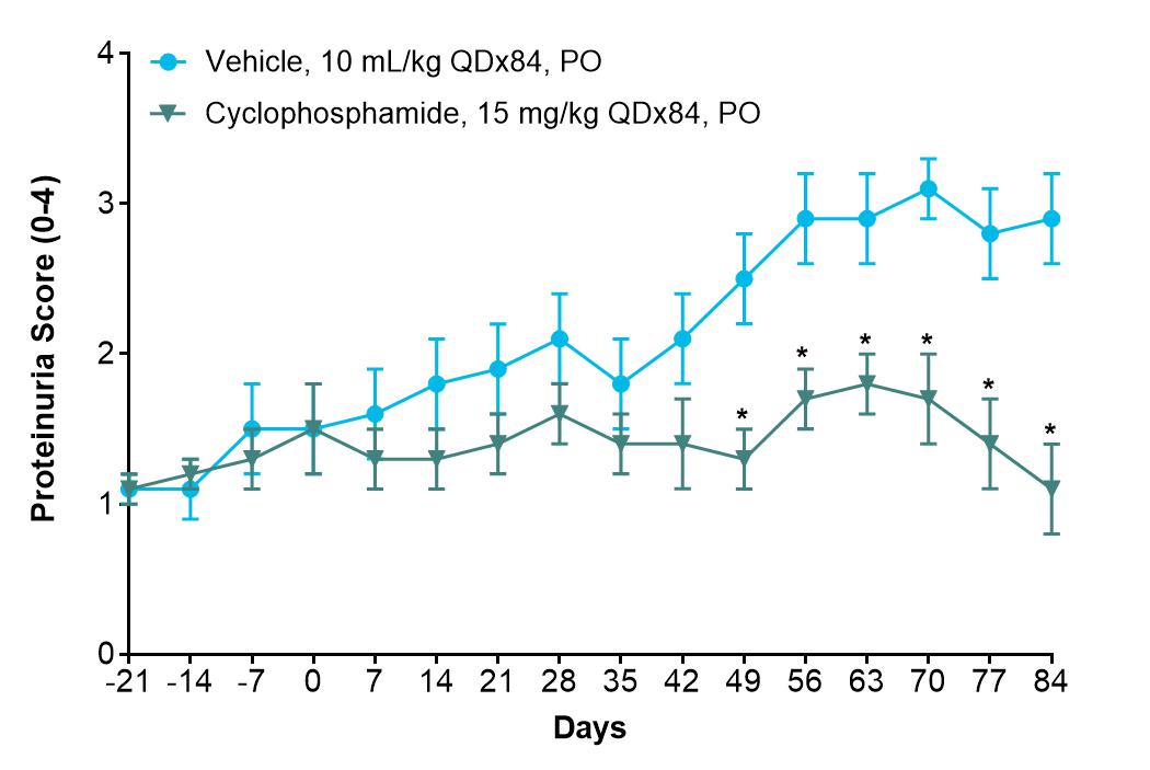
Groups of 10 female MRL/lpr mice purchased from Jackson Laboratory at 10-12 weeks of age are used. Female MRL/lpr mice of 10-12 weeks of age are used in the study. The urine is screened three times per week for proteinuria, and the level of proteinuria is graded according to the manufacturer’s instructions. Mice are randomly assigned to different treatment groups, each with 10 animals, based on the level of proteinuria as well as the body weight after there is kidney damage (glomerulonephritis) as evidenced by the presence of proteinuria of ≥30 mg/dl on two occasions two days apart at around 10-12 weeks of age. After kidney damage, vehicle and test articles are administered orally (PO) once daily (QD) for 12 weeks. The positive control, cyclophosphamide (CTX) at 15 mg/kg is given orally once a day for 12 weeks. Body weight, proteinuria score, and macroscopic skin lesions are recorded once a week, and mortality is observed once daily. Serum blood urea nitrogen (BUN) and creatinine are measured by optimized UV method with an automatic analyzer every 4 weeks after dosing. Titers of total IgG and anti-dsDNA antibodies in serum are also detected by ELISA kit every 4 weeks thereafter. The animals are sacrificed at the end of Week 24, and kidneys, spleen, and mesenteric lymph nodes are collected and weighed. Kidneys are paraffin-embedded and stained with hematoxylin and eosin for histopathology (optional). Data are expressed as a mean ± SEM. Two-way ANOVA followed by Bonferroni’s te
CRITERIA FOR SIGNIFICANCE
Two-way ANOVA followed by Bonferroni’s test is applied for comparison between vehicle and treated groups. p<0.05 is considered significant.
TURNAROUND TIME
STANDARD
120 Days
CLINICAL RELEVANCE
THERAPEUTIC AREA
Autoimmune
ADDITIONAL INFORMATION
BRAND
PDS
TESTING LOCATION
Taiwan - Taipei
OTHER INFORMATION
We will readily accommodate client-specified alterations. These studies are performed at our AAALAC accredited BSL-2 laboratory in Taipei, Taiwan. All aspects of this work are performed in general accordance with the Guide for the Care and Use of laboratory animals (National Academy Press, Washington, DC, 2011). The study protocol was approved by the Pharmacology Discovery Services IACUC and is performed with the oversight of veterinarians to assure the humane treatment of laboratory animals.
ITEM DOES NOT EXIST
© Copyright 2026, Pharmacology Discovery Services – Privacy Policy | Cookie Notice | Cookie Preferences